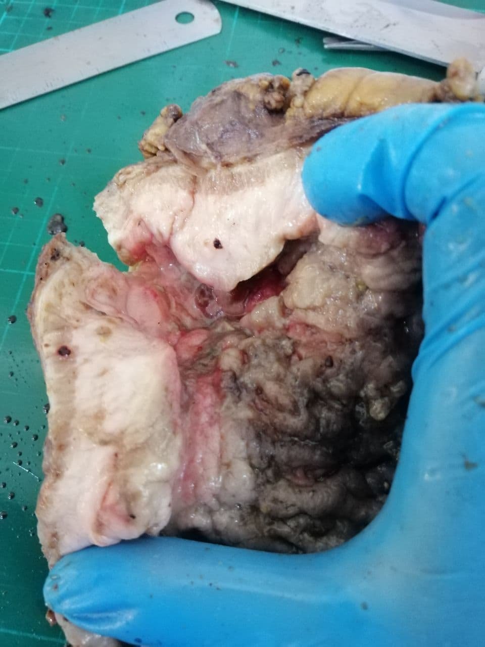
Perineural and Lymph Node Tumor Invasion in a Gastric Carcinoma
7 comments
Perineural and Lymph Node Invasion in a Gastric Carcinoma
Invasion of the perineural and intraneural area signifies poor prognosis and rapid spread towards other organs. Usually, gastric carcinomas are discovered late or as incidental finding which makes them hard to manage when they actually start showing signs in patient. It’s no surprise to see some lymph nodes turned out positive for tumor involvement upon further dissection.
The case was from a mixed gastric carcinoma showing tubulopapillary and discohesive components. I didn’t take a photomicrograph of the tubular to papillary gland component. If you're curious about the grossing of the specimen, here's a link to a different case of poorly cohesive carcinoma of the stomach.

Showing you the typical gross image of these specimens. The photomicrographs below came from a different case but still a stomach specimen.
Taken at Scanner View (40X)

You can see a strand of nerve in a sagittal section surrounded by discohesive neoplastic cells. This was taken from a case of a poorly cohesive carcinoma of the stomach. Discohesive neoplastic cells can come in signet-ring looking type to non-signet ring looking (shown in this case).
Taken at Low Power View (100x)

Taken at High Power View (400x)

When a tumor metastasizes to different parts, they vaguely take form to something as close to the main tumor. That means if they look like glands from the main source, they will attempt to form like glands on any other parts they spread to. The morphology during the spread also gives us a clue where the tumor may have come from like signet-ring cells present in the ovary may have come from the colorectal or gastric part.
Here is what metastasis to regional lymph nodes look like (taken from the same case). You can see the tumor cells (red) invading the remnants of the normal lymph node architecture (green). The neoplastic cells try to form discohesive sheets to tubular glands though it’s not as prominent on the images below.
Taken at Low Power View (100x)

Taken at Low Power View (100x)

We look for several lymph nodes in the specimen and see how many are positive then how large are the tumor deposits. This affects the prognostic staging of the patient. We use the TNM classification for staging.
If you made it this far reading, thank you for your time.
Posted with STEMGeeks


Comments