Some Pictures of Anaplastic Large Cell Lymphoma Revisited
10 comments
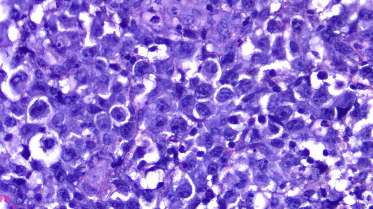
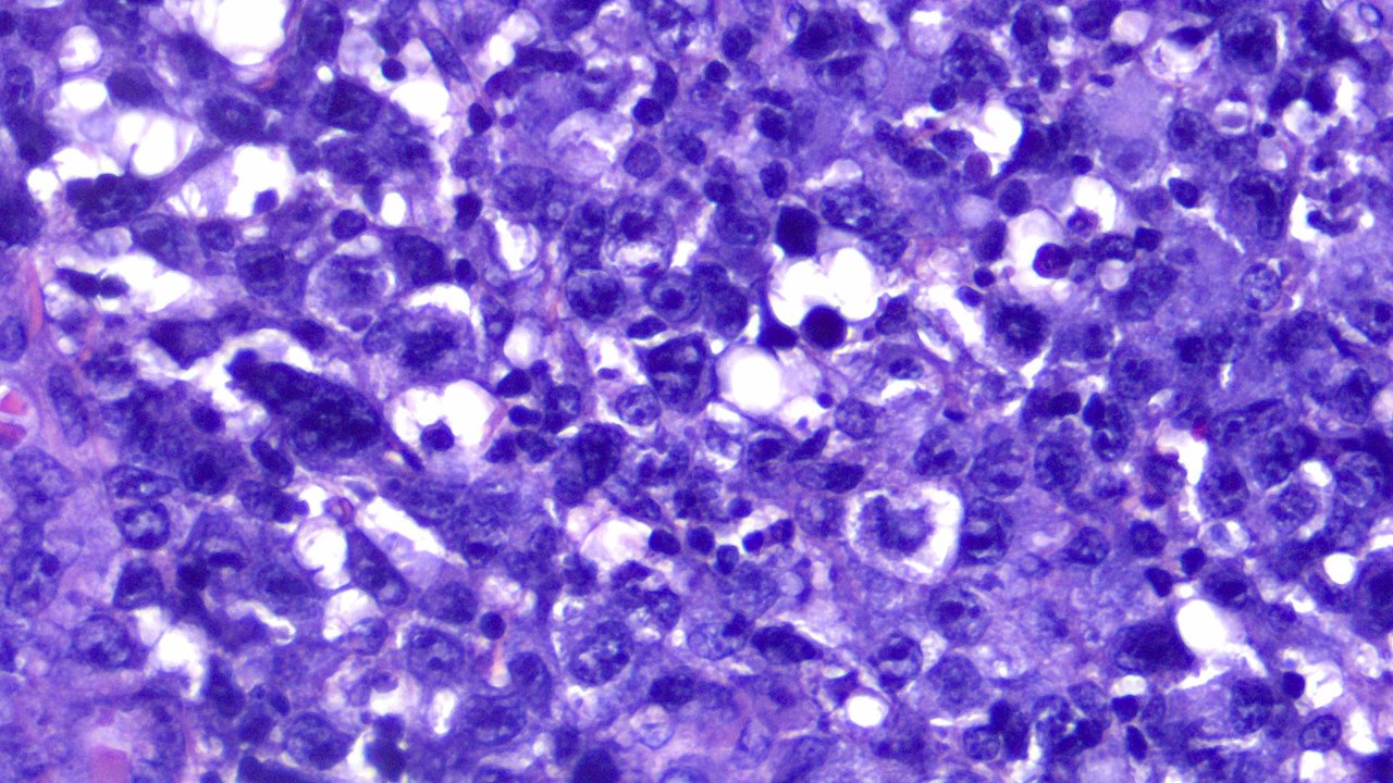
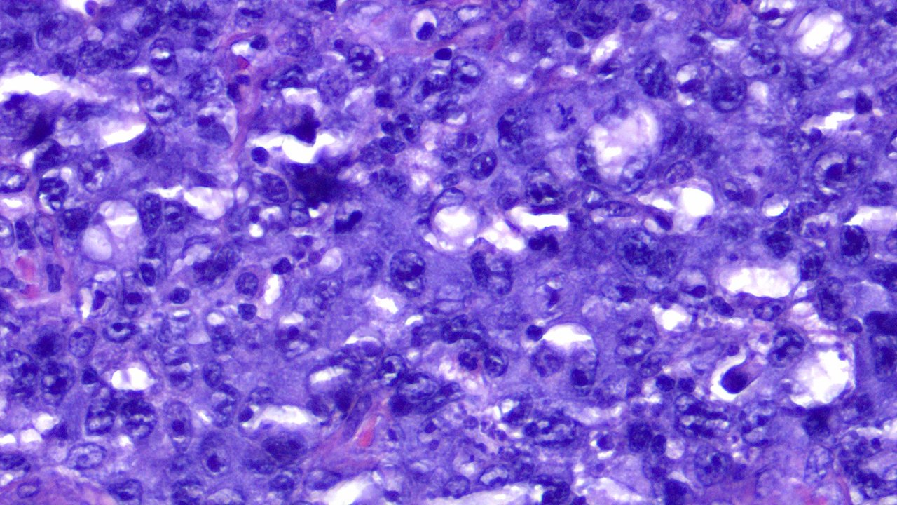
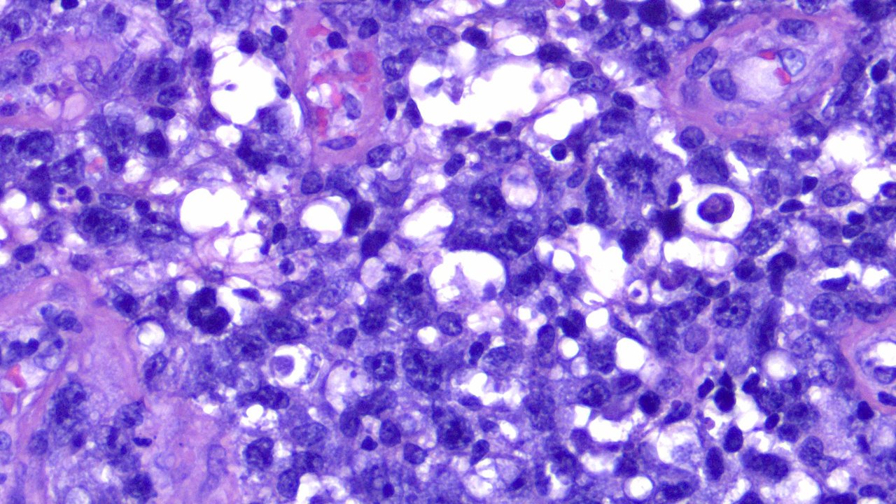
This is the same case of Anaplastic Large Cell Lymphoma previously shared here with different sets of images. I got to revisit the case after the physician requested a new set of images for their case report series for publishing.
These were all taken from a 400x magnification view. Everything you see here are sheets of tumor cells that are discohesive with neighboring cells. Nothing looks ordinary, there's no regularity on their membranes, some cells are actively dividing, there's no clear boundaries between cells and the hyperchromatic nuclear membranes tell it's actively producing something to prolong their lives and replicate.


A lot about the case has already been said on the previous post. I'm just sharing these pics as it's also one of the cases that I'll never forget in my career under Pathology Training given how much time and resources I spent just to facilitate some closure over this.
In a resource limited setting, I sometimes pay for the expenses the patient incurs out of pocket without expecting any reimbursement. If you think I'm being generous, that's one way of looking at it but to me, I'm just doing it because I can, have the choice and probably sleep better at night for selfish reasons that had good outcomes. The patient already expired, not sure if I mentioned this in the previous post.
I'm not really sure if I'll be posting on this account again in the future. Maybe it would just be random science stuff I found online as I had the idea of just using this account for sharing interesting finds at work. I'm going to be unemployed next year and will reevaluate on whether I want to come back to Pathology training or shift to another field of specialty in medicine. Probably my last post here until further notice.
If you made it this far reading, thank you for your time.
Posted using STEMGeeks
Comments