blowing the lid on a couple of fungi
7 comments
this is my contribution to #FungiFriday by @ewkaw
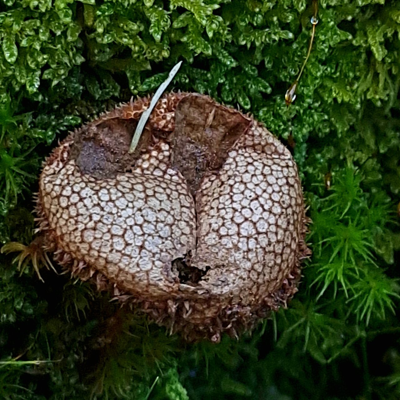
Lycoperdon echinatum spiny puffball
when it's young. this puffball is a sphere covered with spines. it is edible as long as it is completely white inside with an even consistency and no trace of any gills. being white inside indicates that the fungi have not yet developed any spores. unlike other fungi with gills or pores puffballs produce all their spores internally and keep them inside until mature and dry. if i had found this when it was young i may have picked it to eat. anyway, now it is old and dried out and looks like a handsome sack with a netlike covering. the net design marks where the spines used to be. the top of this one is missing but it has not yet released many spores. what usually happens is the puffball forms an aperture or opening on the top. then when something strikes it, most likely raindrops, it releases spores in what looks like small clouds of dust. now a raindrop may not produce much of a cloud of spore powder but a gentle tap with a finger will!
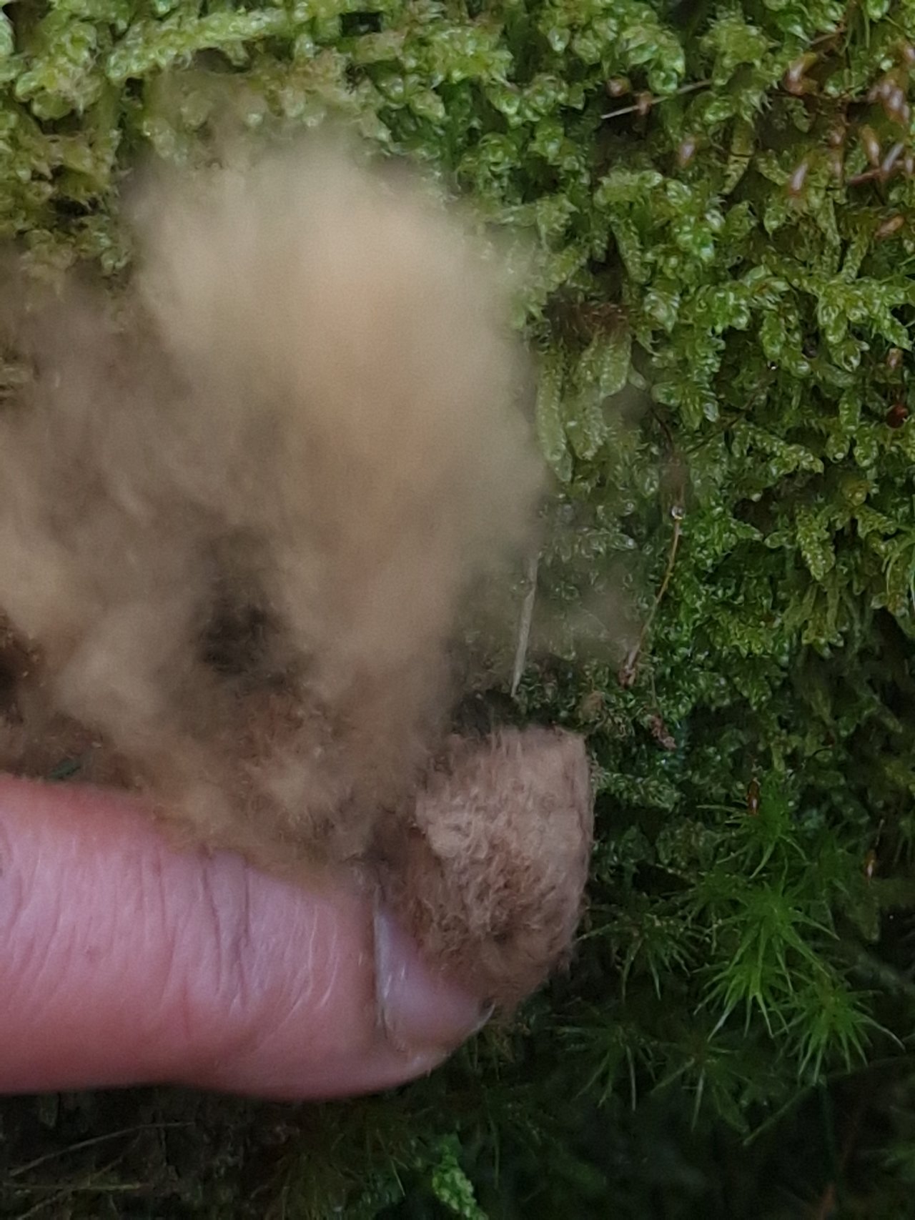
it's very fun to poke them and you can repeat it several times. since this is their way of reproducing they don't mind at all. in fact a good poke is exactly what they need and want most in life 😂😂😂😂. it spreads their spores far wider than some mere raindrops could.
it can be difficult to identify dried puffballs but the net design is a giveaway for this species. the brown cloud contributes as a confirmation. other puffballs may have brown, purple or green spores
there are many aspects i find fascinating about fungi. not the least is the way they distribute their spores. typically they have gills or pores under their caps and the spores are released and simply fall down to the ground. i like how puffballs take a completely different approach. and there are many different species of fungi with totally different and often unique strategies and methods.
for instance Paragyromitra infula elfin saddle or hooded false morel
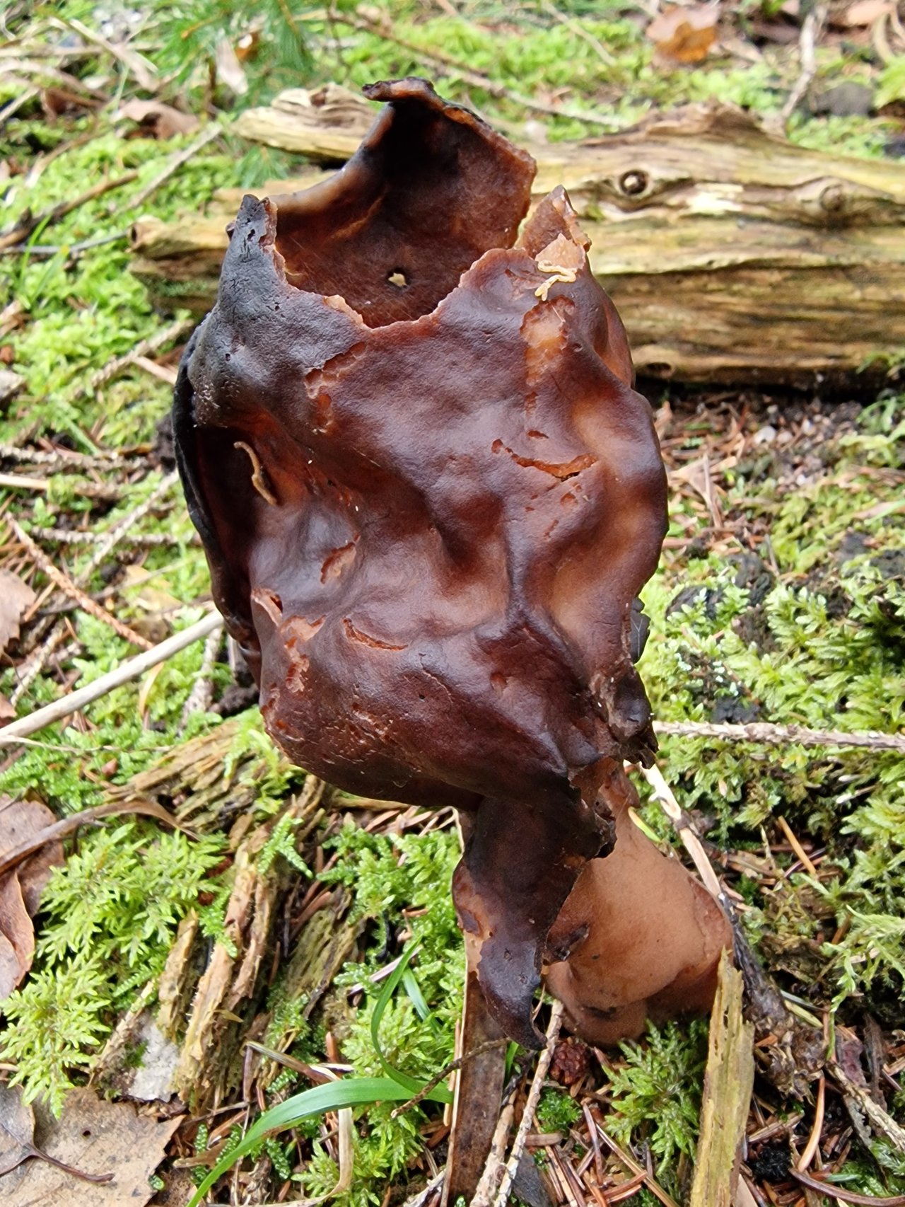
this one has also lost it's top. i don't know how it happened but normally the top is sealed. because i don't pick these i have never seen inside one.
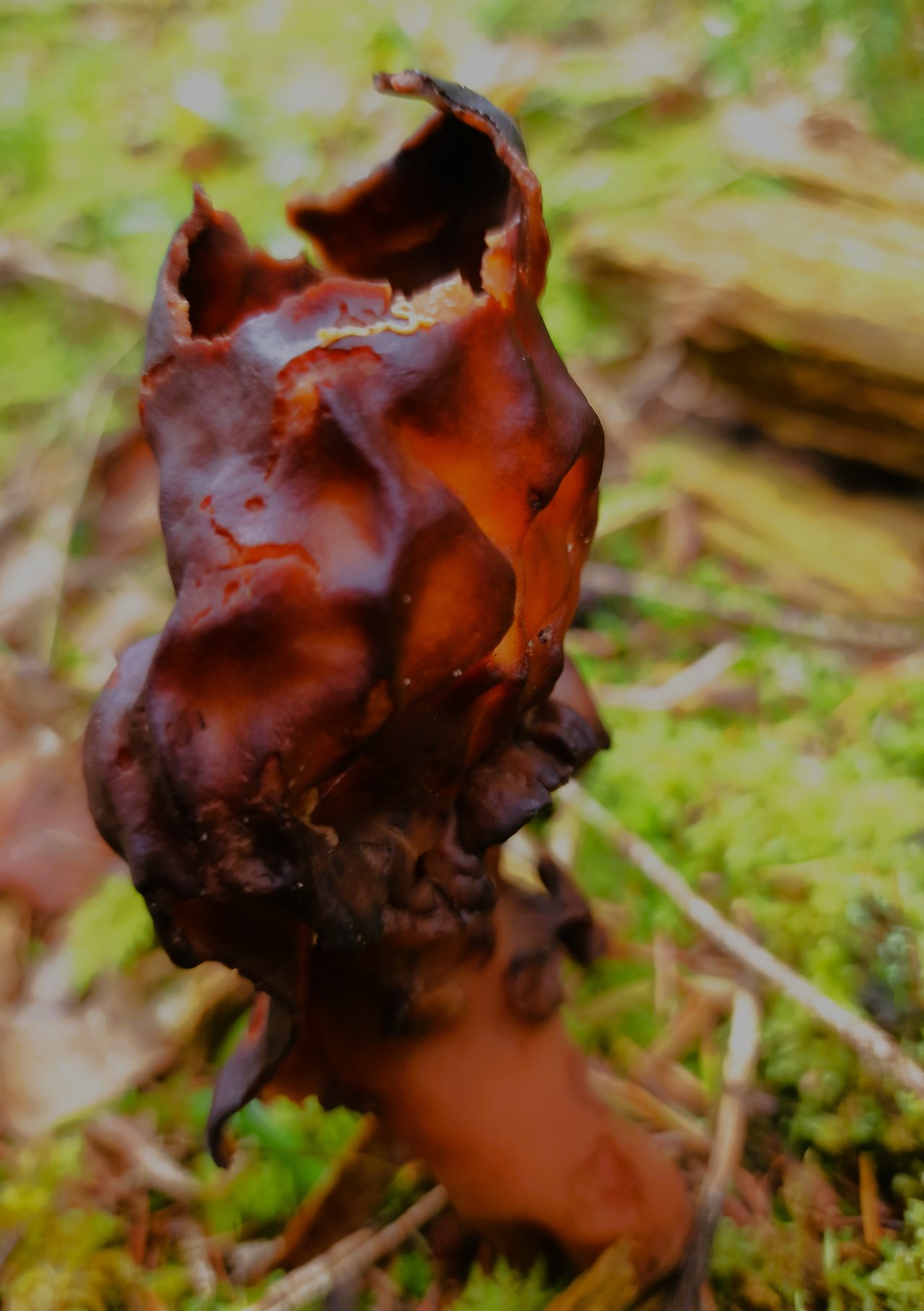
now that i could see inside i realized it was hollow with very thin somewhat translucent walls. without the top in place light enters inside and shines through the thinnest parts which brings out paler shades of the otherwise dark reddish brown.
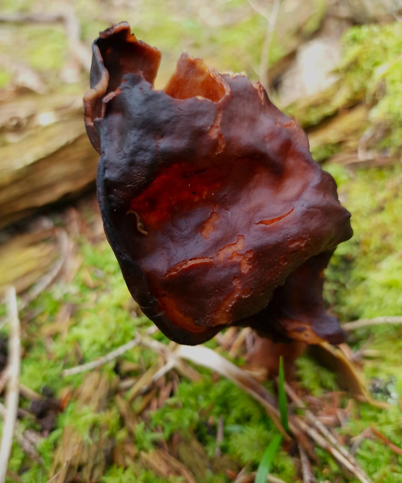
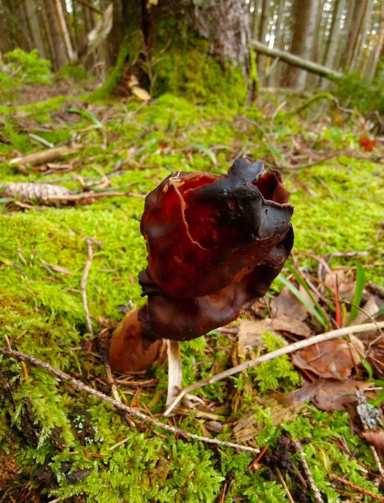
even with it's head on straight this one wouldn't win a fungi beauty contest or an election without cheating
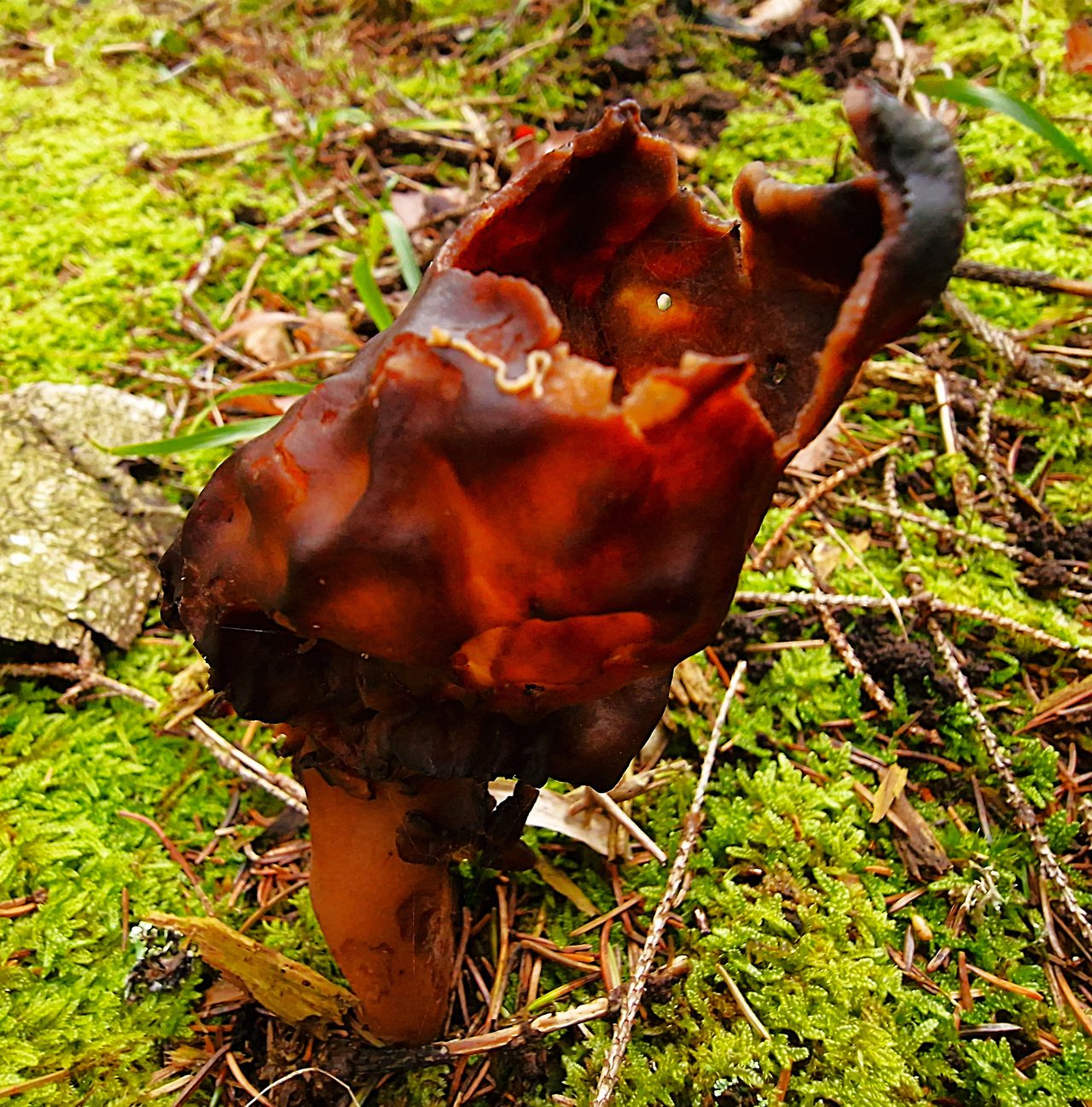
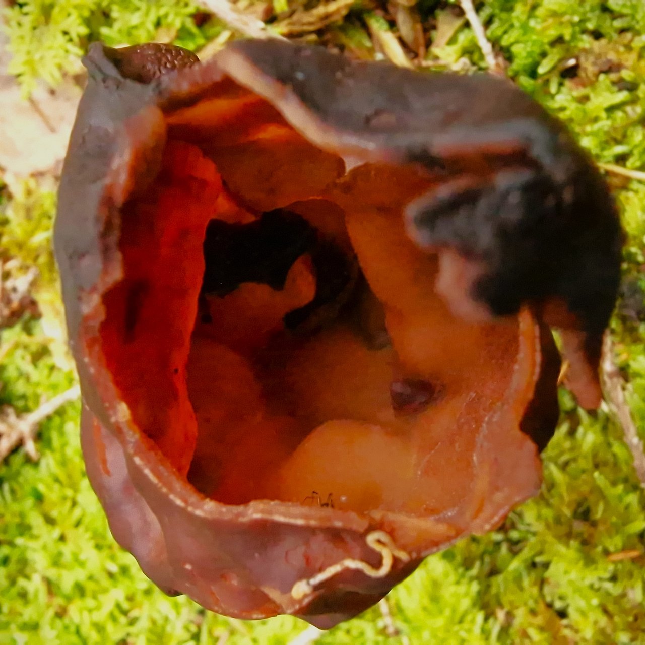
but at least i figured if i look inside i might be able to see some signs of spores or a clue as to how it disburses them. sadly, i couldn't make heads or tails of what i saw. so i went on the internet in hopes of learning something.
Wikipedia states:
Similar to a cleistothecium, a gymnothecium is a completely enclosed structure containing globose or pear-shaped, deliquescent asci. However, unlike the cleistothecium, the peridial wall of a gymnothecium consists of a loosely woven "tuft" of hyphae, often ornamented with elaborate coils or spines.
and elsewhere wikipedia eloquently elucidates:
Ascospores are ellipsoidal in shape, hyaline, smooth, thin-walled, with dimensions of 17–22 by 7–9 μm.[14] They are also biguttulate, containing two large oil droplets at either end. The spore-producing cells, the asci, are roughly cylindrical, eight-spored, operculate (opening by an apical lid to discharge the spores) and have dimensions of 200–350 by 12–17 |μm
Ah, operculate that explains it. i should have known that the ascospores are both hyaline and biguttulate so i'm glad i finally got that cleared up as well. now i'm proud to be able to blow the lid for all Hiveans that these peculiar drack looking organisms manage to spread their spores because their asci are operculate!
it's amazing how they developed that method. absolutely brilliant. the more i think about it, the more astounding fungi are. it blows my mind.
Comments