Malignant Cells in a Thyroid Aspirate
8 comments
Sharing some images I got from the thyroid aspirate specimen taken from a 62 year old female. We called this case as malignant based on the overt cytologic clues. Because it's only an aspirate done for this sample, we're unable to commit to a definite type of cancer. One needs a tissue sample for a complete histopathologic diagnosis.
Fine needle aspiration biopsies are done to check whether the tumor is malignant or benign. One of the benefits of aspirating samples is it's less invasive and a possible diagnosis between malignant and benign can be made. Though errors can come from poor sampling technique which doesn't rule out carcinoma if the imaging studies done suggests the mass looks malignant.
Anyway, the purpose is here is just appreciating the clues that made us call this case as a positive for malignancy. I usually have a hard time diagnosing thyroid aspirate specimens given the lack of cellularity but this one was a classic textbook appearance on what I'm supposed to look for.

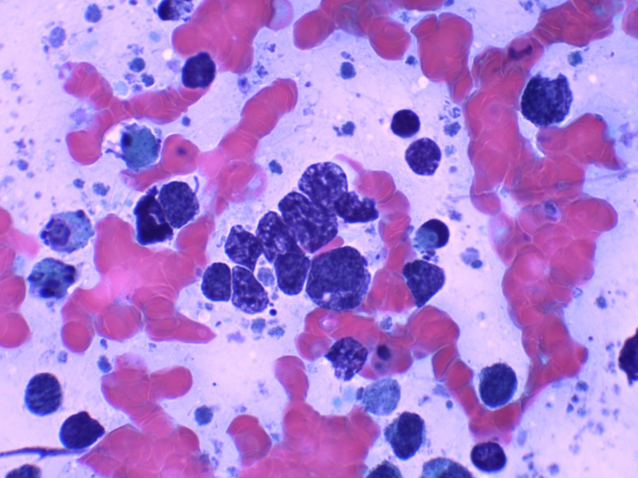
Right at the middle, you'll see a cluster of malignant cells with irregular nuclear membranes instead of the normal uniform small and round nuclei found in benign thyroid follicular cells. You only need to verify that the sample was taken from the thyroid and this image on the microscope to be certain it's malignant.
Pardon the poor quality image and lighting, this was taken by my phone and not the usual camera I use for taking pictomicrographs. You'll also notice the surrounding debris that hint some necrosis which is a common feature for malignant pathologies. This was a bloody aspirate due to the prominence of red blood cells surrounding the tumor cells.

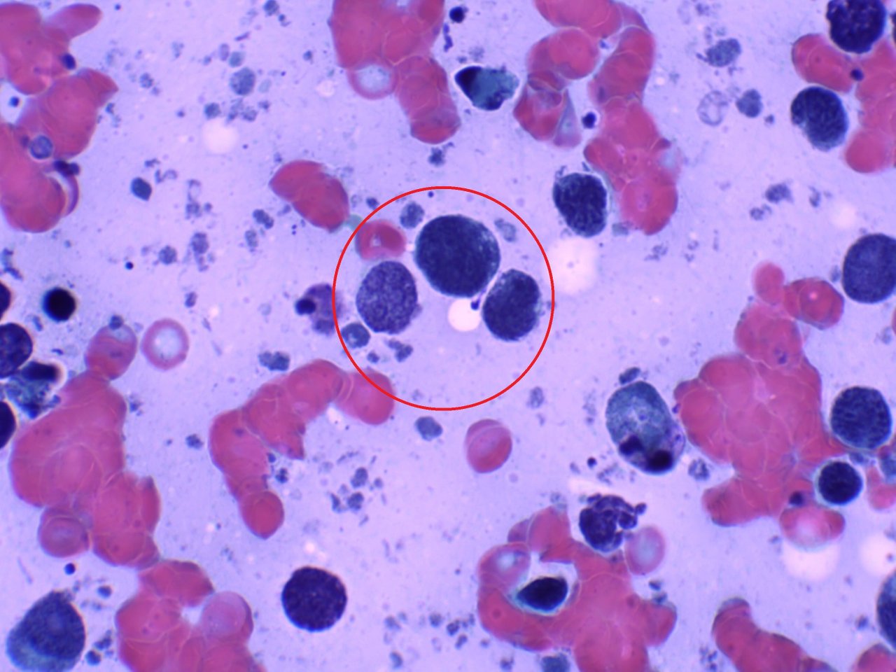
Tumor cells are hyperchromatic (more blue) but they can display different levels of intense staining which can highlight some cellular features like a prominent nucleoli. The one on the left with some eyes and lighter stain shows a fine chromatin pattern which indicates a highly active metabolic process is taking place. Fine chromatin means DNA is let loose for transcribing more proteins but not all tumor cells have fine chromatin.

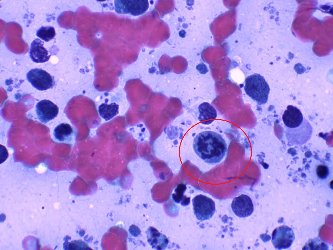
Here is an atypical mitosis taking place. This further adds weight to the conviction of the specimen being malignant.

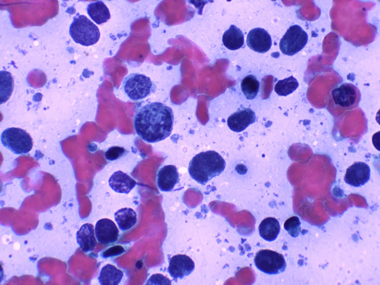
Some of these darker staining cells, especially the small ones may just be lymphocytes reacting to the tumor cells. I get fascinated whenever I see specimens like these given how cool they look live. But at the end of the day, this learning experience is at the cost of someone else's misery as a diagnosis of malignancy and further workup is indicated in the report.
If you made it this far reading, thank you for your time.
Posted with STEMGeeks
Comments