Fine Needle Aspiration Biopsy and Tissue Biopsy
5 comments
Fine needle aspiration biopsy (FNAB) is poking a needle on the suspected mass and then putting the contents aspirated on the slide for processing and cytology to diagnose. It's not the best method to diagnose as a tissue biopsy is still better. It's flaws include operator technique when the case is malignant but you get benign findings because they missed the tumor.
But this method is less invasive because this lets the surgeon decide whether to proceed cutting up the mass open or leave it for observation. Sharing an image previously mentioned on papillary thyroid carcinoma post
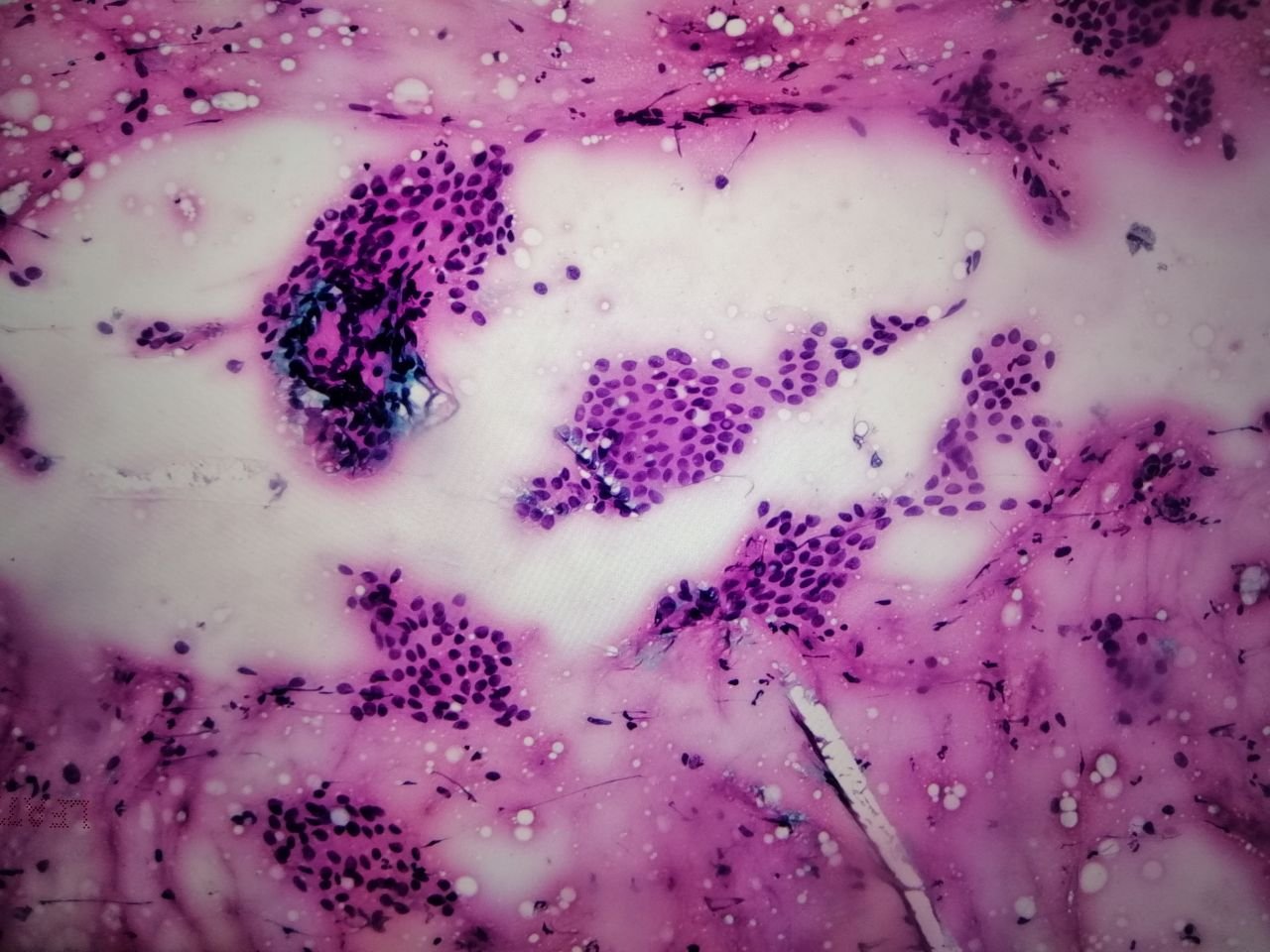
Sharing you some pics from my previous cases. The cytologic smears below came from a 48 female who presented with a neck mass. That's just the information we get on the requests especially if the sample came from far flung institutions. My pet peeve is reading requests with limited data because that can stall the diagnosis.

This is the tissue sections on slides diagnosed with Papillary Thyroid Carcinoma. But below is a case suspected of PTC after FNAB. The images were from my phone facing an LCD monitor attached to the microscope. I'm maximizing the use of the microscope since the vendor left it at the office for demonstration for only a month.
This is the low power view.

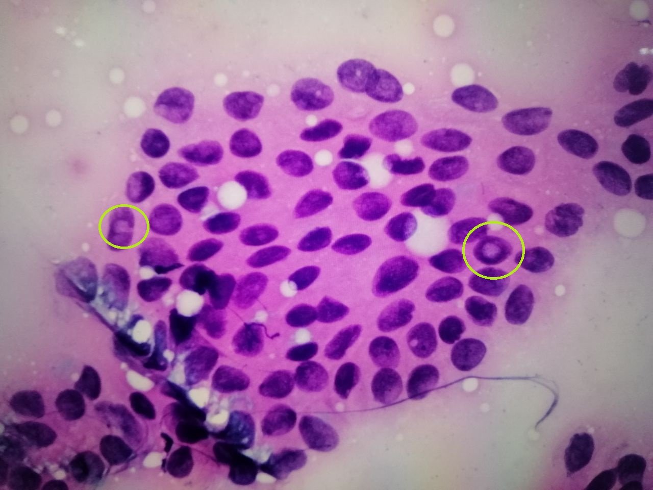
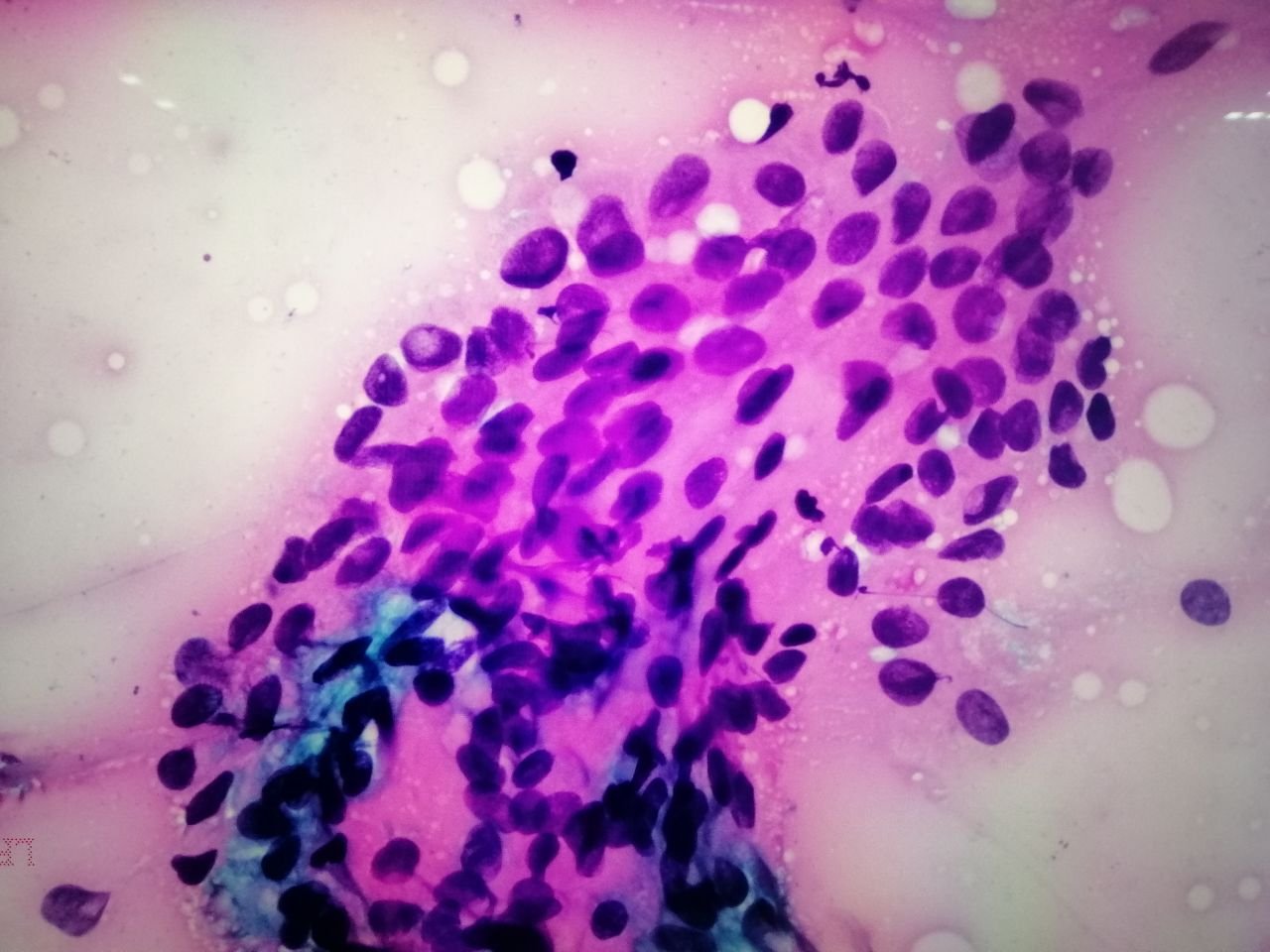
The High power view:
I've highlighted the suspicious cell. Well most of the cells below are already suspicious. I'm looking for cells that have a prominent nucleoli (green circles), they look ovoid or grooved, irregular nuclear membrane and pale.


Because the morphology of these cells don't look uniform and and the pseudoinclusions present suggest the case is PTC. But final diagnosis will still depend on tissue resection. Now the surgeon has a basis to perform surgery.
If you made it this far reading, thank you for your time.
Posted with STEMGeeks
Comments